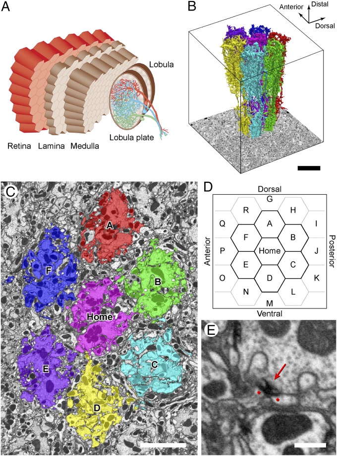
Fig. 1.
Seven-column connectome reconstruction. (A) Overview of the optic lobe of Drosophila, showing the repeating retinotopic architecture of successive neuropils. Modified with permission from ref. 24. (B) Three-dimensional reconstruction of modular medulla cell types in each of seven reconstructed columns. (C) Transverse section in distal medulla stratum M1. Columns (Home and A–F) are colored to conform to B. (D) Plot of a column array. The central Home column is surrounded by its six neighboring columns A–F and 12 more in the outer ring. (E) Focused-ion beam milling (FIB) electron micrograph of neurite profiles with a presynaptic T-bar ribbon (arrow) and two juxtaposed dendrites with PSDs, revealed by membrane densities (dots). (Scale bars: B, 10 μm; C, 5 μm; E, 500 nm.)

