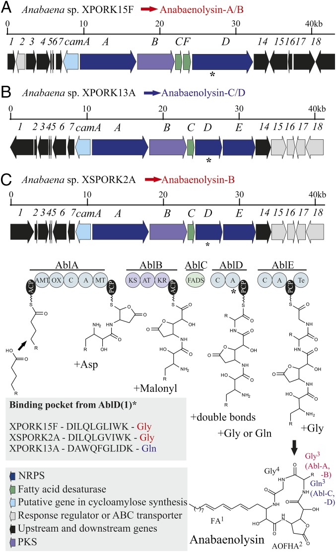
Fig. 4.
Putative anabaenolysin gene clusters from Anabaena spp. XPORK15F (A), XPORK13A (B), and XSPORK2A (C). The main variant of anabaenolysin (A or B in red, C and D in blue) produced per strain is indicated in the figure. The main difference between the variants is the glycine (A/B) or glutamine (C/D) in position 3. The signature of the binding pockets from the adenylation domain of AblD(1) appears in the figure, and an asterisk (*) denotes the AblD(1) region in the gene cluster.

