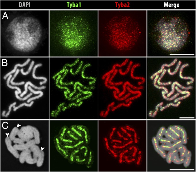
Fig. 1.
FISH localization of Tyba1 and -2 in R. pubera. (A) Hybridization signals of both Tyba subfamilies in an interphase nucleus show a genome-wide dot-like labeling. (B) Prometaphase chromosomes show a line-like but dispersed labeling on the poleward surface of each chromatid and (C) a distinct labeling along the centromere groove of both sister chromatids during metaphase. Arrowheads in C indicate grooves. (Scale bars: 5 μm.)

