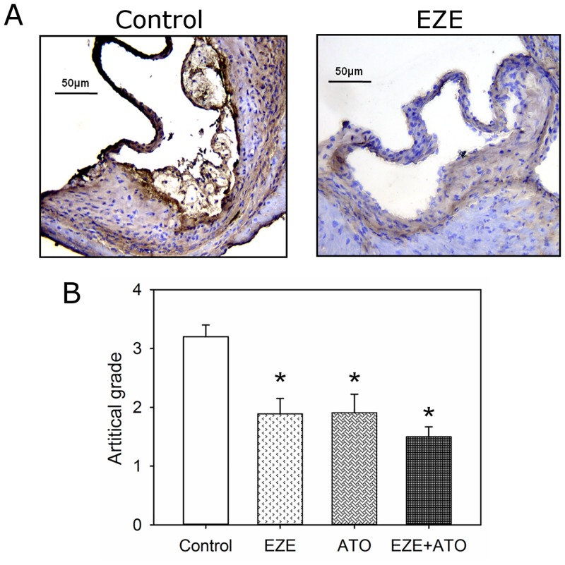
Fig 5. Drugs suppressed macrophage accumulation in atherosclerotic lesions.
Macrophage (CD68) was detected by immunostaining on cross-sections (10 μm thick) of aortic roots. According to artificial grades of positive immunostaining (as shown in S5 Fig), macrophage contents were assessed semiquantitatively. (A) Representative tissue sections by immunostaining. Left panel indicated the mice in control group, whereas right panel indicated the mice in ezetimibe group (EZE). (B) Artificial grades of macrophage contents in lesions of the mice. Histobars represent means, and error bars represent SEM. N = 9, 8, 11 and 11 for vehicle (Control), ezetimibe (EZE), atorvastatin (ATO), and combination (EZE+ATO) groups, respectively. Statistical analysis was performed using one way repeated measure ANOVA. * P<0.001, compared with vehicle.

