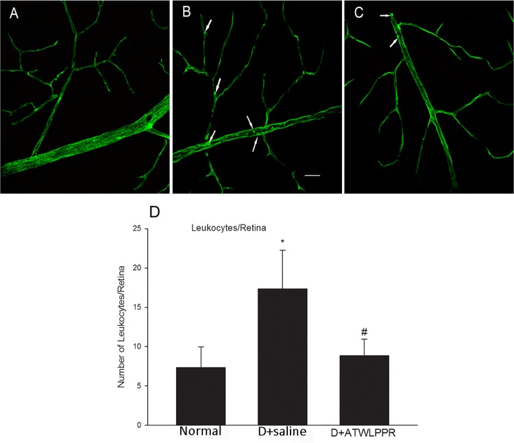
Fig 4. Treatment with ATWLPPR reduced the leukocyte attachment.
Con A perfusion retinas show attached leukocytes (arrows) within the vasculature of diabetic retinas. Representative retina flat-mount images from (A) Normal, (B) D+saline and (C) D+ATWLPPR groups. (D) Quantitative analysis of leukocyte adhesion. Scale bar = 40 mm. *P < 0.05, versus the Normal group; #P < 0.05, versus the D+saline group. D, Diabetic.

