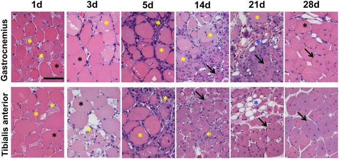
Fig 2. Morphometric analysis.
Representative Hematoxilin/Eosin stained sections of Gastrocnemius (top) and Tibialis anterior muscles (bottom) following ischemia. Calibration bar = 50 μm. Black asterisks indicate healthy myofibers with peripheral nuclei. Yellow asterisks indicate damaged tissue, including necrotic myofibers and inflammatory infiltrating cells. Necrotic myofibers were identified by differential eosin staining and presence of infiltrating cells inside the fiber. Blue asterisks indicate adipose cells. Black arrows indicate regenerating myofibers characterized by the central nucleus/i.

