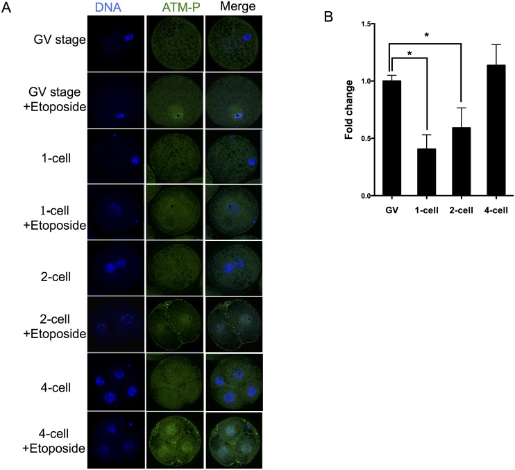
Fig 5. Phosphorylated ATM in the oocytes and preimplantation-stage embryos.
(A) Immunolabeling for active ATM at 100 μg/mL etoposide treatment. (B) Fold change in ATM-p was determined using the formula F-F0/FGV-F0, where F is the mean value of each etoposide treatment, and F0 is the control value where no etoposide is used (0 μg/mL). FGV is the value of GV stage exposure to 100 μg/mL etoposide. This formula was used to be able to disregard nonspecific staining, which allows for a better comparison of the different cell types.

