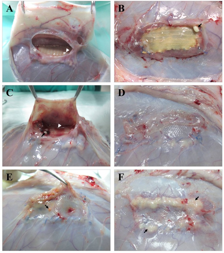
Fig 4. Macroscopic findings.
Appearance of the different meshes after 14 days of implantation and Sa contamination. (A, B) DM+ implants showing thick fibrous encapsulation (*), seroma formation (▶) and the presence of dispersed purulent material (→) associated with the mesh anchorage, while the main body of the implant remains clean. (C, D) PP + CHX implants showing similar behavior to the previous ones, with the exceptions of a more intense vascularization and total mesh integration into the host tissue. (E, F) PP + allicin-CHX implants show large amounts of purulent material (→) covering different areas of the implant surface.

