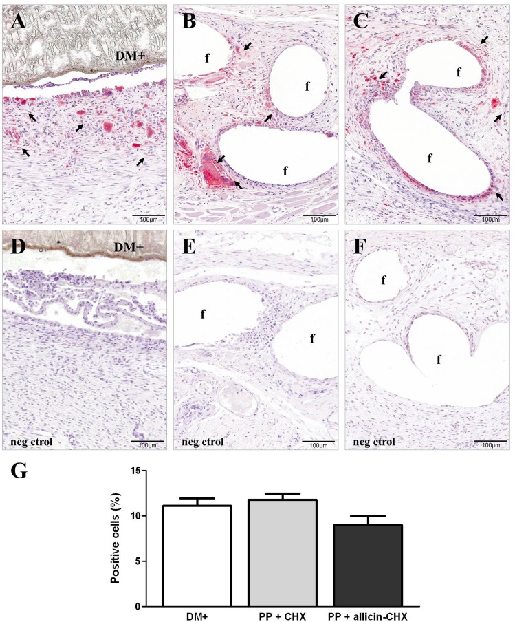
Fig 7. Foreign-body reaction.
RAM-11 immunostaining (x200) of the (A) DM+, (B) PP + CHX, and (C) PP + allicin-CHX implants showing the presence of labeled macrophages (→) in the neoformed tissue. (D-F) Negative controls of the DM+ (D), PP + CHX (E) and PP + allicin-CHX (F) implants showing no immunostaining. (G) Positive cell percentages recorded after 14 days of implant. The results are expressed as the mean ± standard error of the mean for the total of micrographs counted (7 specimens per study group, 10 micrographs per specimen). The lower percentage of RAM-11 positive cells recorded for the PP + allicin-CHX implants was not statistically significant.

