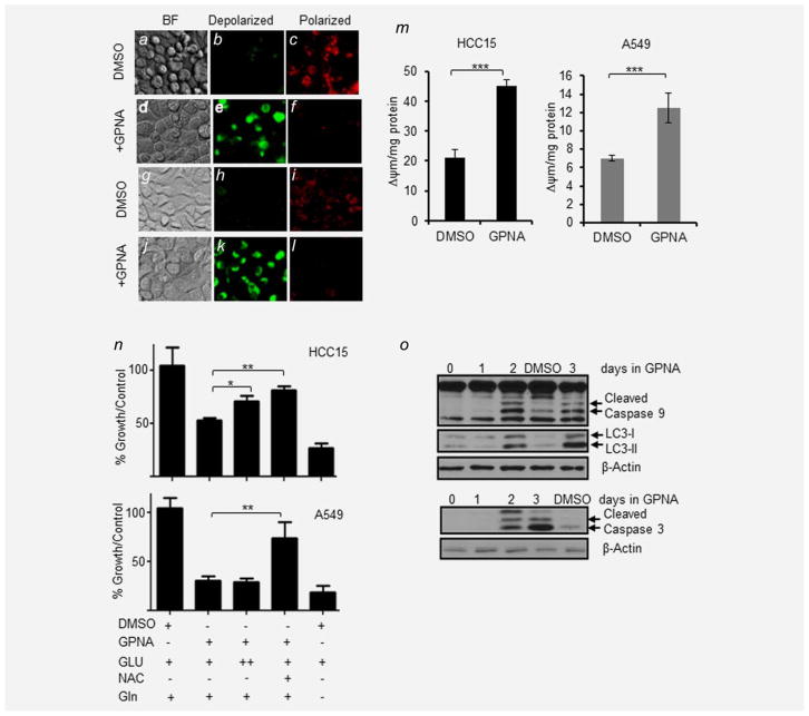
Figure 4.
Targeting SLC1A5 induces autophagy and apoptosis. JC-1 dye was used to measure mitochondrial potential (Δψm) in live HCC15 and A549 cells after 24 hr of 500 μM GPNA or DMSO (0.01%, v/v). (a–c) HCC15 treated with DMSO (0.01%), where (a) is bright field image of HCC15 cells, (b) is a depiction of depolarized mitochondria shown as green fluoresce of JC1 monomers and (c) shows polarized mitochondria as red fluorescence of JC1 aggregates. (d–f) HCC15 treated with GPNA in the same sequence. (g–i) A549 treated with DMSO (0.01%) and (j–l) A549 treated with GPNA. (m) Effect of 500 μM GPNA on mitochondrial potential (Δψm) expressed as the ratio of green/red in HCC15 and A549. (n) Effect of GPNA, glutamate (GLU), glutamine (Gln) and N-acetylcysteine (NAC) on A549 and HCC15 (both high expressing SLC1A5 lines). All data are the averages ±SEM of at least three separate determinations (*p <0.05, **p <0.005). ++, tenfold higher concentration of glutamate was added. (o) Effect of 500 μM GPNA on induction of autophagy (decrease in LC3I and increase of LC3-II) and intrinsic apoptotic markers (Caspase 9 and 3) in a time-dependent manner in HCC15 cells.

