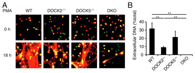
FIGURE 5.
DKO neutrophils exhibit a severe defect in NET formation. (A) Following stimulation with PMA (20 nM) for 18 h at 37°C, WT, DOCK2−/−, DOCK5−/−, and DKO neutrophils were stained with Sytox Green and Sytox Orange for visualization of NETs (original magnification ×200). (B) NET formation was quantitatively compared among PMA (20 nM)-stimulated neutrophils from WT, DOCK2−/−, DOCK5−/−, and DKO mice. Data are indicated as means ± SEM of five separate experiments. **p < 0.01.

