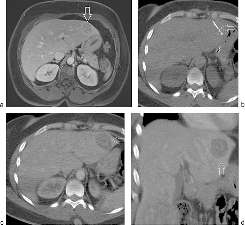
Fig. 1.

(a) Diagnostic MRI of the liver with intravenous contrast showing the mass in the lateral segment of the left hepatic lobe (arrow). (b) CT imaging showing a 15-cm Thermosphere ablation probe (arrow) in the lesion in the lateral segment of the left hepatic lobe. (c–d) Postablation CT scan with intravenous contrast showing the zone of ablation (arrow) in the axial and coronal planes.
