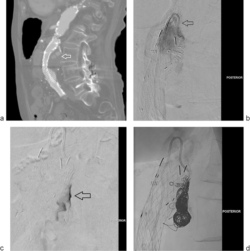
Fig. 4.

(a) Sagittal reconstruction from a CTA performed in a patient with history of endovascular repair of an abdominal aortic aneurysm. The image shows a type-I endoleak that fills the posterior aspect of the aneurysmal sac (arrow). (b) Selected image of a digital subtraction arteriogram performed in a true lateral view shows contrast injection through a reverse-curve catheter (arrow) aimed posteriorly, toward the expected site of the endoleak. The endoleak is clearly demonstrated. (c) Selected image of a digital subtraction arteriogram performed through a microcatheter within the endoleak sac. The endoleak sac is clearly opacified (arrow). No feeding collaterals are identified. (d) Selected spot film obtained after hydrogel-coated coil packing of the endoleak cavity.
