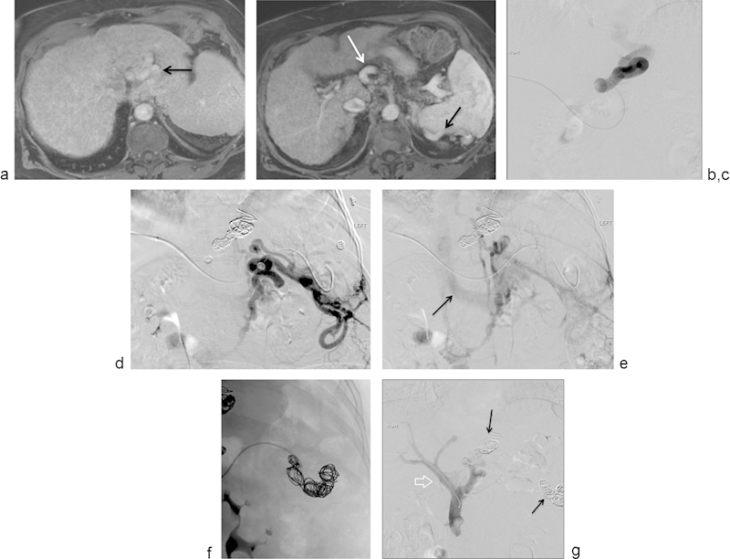
Fig. 6.

(a) Selected axial image of a contrast-enhanced MRI showing large vascular structures posterior to the left lobe of the liver, corresponding to large esophageal varices (arrow). (b) Selected axial image of a contrast-enhanced MRI showing large vascular structures anterior to the celiac trunk (white arrow) and posterior to the spleen (black arrow). These venous structures were thought to be part of a large splenorenal shunt. (c) Selected image from a digital subtraction angiogram showing injection into a pocket of large esophageal varices. No flow into the main portal vein is demonstrated. (d) Selected image from a digital subtraction angiogram showing injection into a large, complex, splenorenal shunt. (e) Selected delayed image from a digital subtraction angiogram after injection of contrast into a large, complex, splenorenal shunt demonstrates opacification of the left renal vein (arrow), draining into the inferior vena cava. (f) Spot film that shows the process of embolization of the large splenorenal shunt with hydrogel-coated coils. (g) Selected image from a digital subtraction direct portogram showing successful coil embolization of the portosystemic shunts from the left gastric vein and from the splenorenal shunt. There is now antegrade flow into the main portal vein (arrow) and intrahepatic branches. Patient's encephalopathy improved significantly after this intervention.
