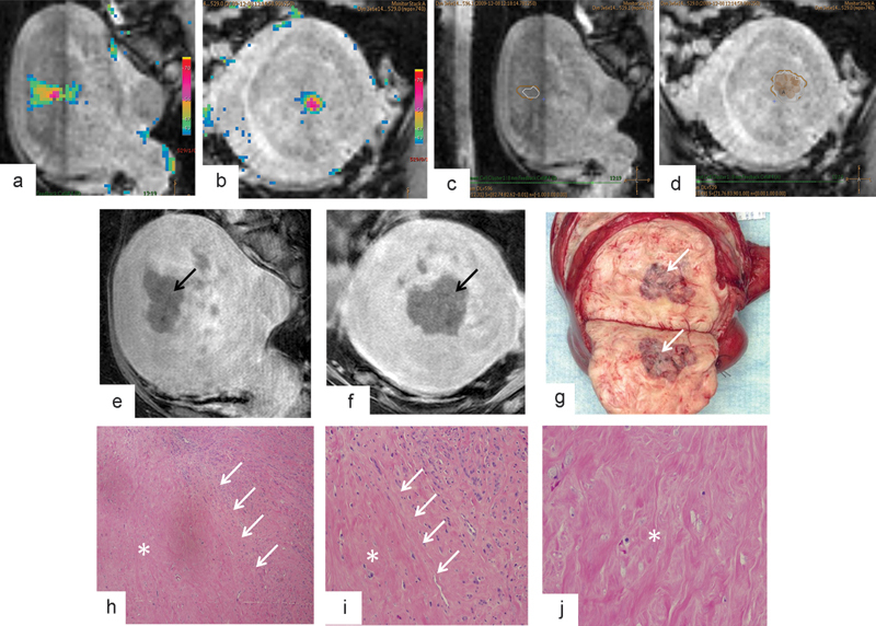
Fig. 4.

Intraprocedural MR-guided HIFU monitoring and MR imaging and histopathologic findings after leiomyoma ablation with MR-guided HIFU. (a–d) Graphic user interface displays multiplanar three-dimensional T2-weighted imaging and overlaid temperature maps (a–b) and overlaid thermal dose estimates (c–d) during sonication of an anterior intramural leiomyoma within the body of the uterus. Accumulated thermal dose information in the treated volume is displayed at the end of each sonication as a thermal dose estimate. These thermal doses are reported in CEM43, with 30 CEM43 (beige polygon, c–d) corresponding to onset of tissue alteration and 240 CEM43 (white polygon, c–d) representing predicted territory of complete necrosis. Both 30 CEM43 and 240 CEM43 thermal dose estimates are updated after each sonication. (e–f) Sagittal (e) and coronal (f) contrast-enhanced MR images after HIFU show nonenhancing treated region (black arrows). (g) Bivalved gross uterine specimen shows hemorrhagic necrosis in the area of treatment (white arrow). (h–j) Low-magnification (4 × ) histologic images of margin (h), high-magnification (10 × ) images of margin (j), and high-magnification images of the center of the ablation zone (j) confirm necrosis (asterisk) and narrow zone of transition (white arrows) between viable and necrotic HIFU-treated tissue. (Reprinted with permission from Venkatesan et al.43)
