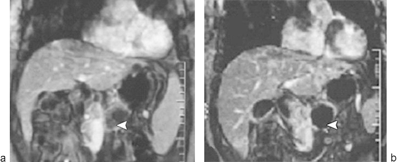
Fig. 8.

Dynamic contrast-enhanced gradient-echo T1-weighted MR images (180/6.0, 90-degree flip angle, 128 × 256 matrix, 10-mm-thick sections, 2-mm intersection gap, one signal acquired, and 18-second acquisition time) obtained with breath holding from a 48-year-old man who underwent high-intensity focused ultrasound ablation for advanced pancreatic cancer. The tumor was 4.5 × 4.5 cm in diameter and located in the body of the pancreas. (a) Image obtained before high-intensity focused ultrasound shows the blood supply in the pancreatic lesion (arrowhead). (b) Image obtained 2 weeks after high-intensity focused ultrasound shows no evidence of contrast enhancement in the treated lesion (arrowhead), which is indicative of complete coagulation necrosis in the pancreatic cancer. (Reprinted with permission from Wu et al.97)
