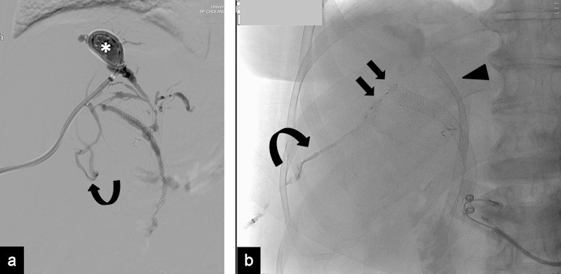
Fig. 11.

(a) Cholangiogram shows a biloma with multiple strictures after biliary drainage in a patient with cholangiocarcinoma and previous metallic stents, presenting with cholangitis (*). Communication with hepatic veins (curved arrow) is also seen. (b) Radiograph shows placement of an additional metallic stent (arrowhead); after stent deployment, two AVP IV plugs were used to close the tract (arrows). Histoacryl glue was then placed to further close the tract and the skin entry site to control ascites leakage. The plugs prevented glue migration into the biliary ducts.
