
Fig. 2.
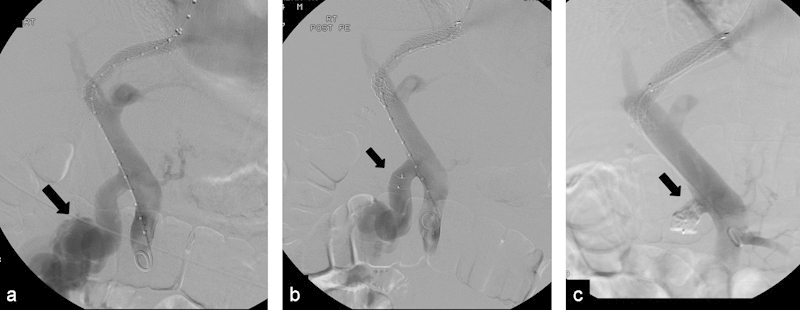
(a) Patient with cirrhosis and portal hypertension with bleeding duodenal varices. Portogram after TIPS shows large portomesenteric shunt (arrow) causing the ectopic varices; an AVP 1 was placed. (b) Patient had recurrent bleeding 2 weeks after initial embolization procedure. Portogram demonstrates that the portomesenteric shunt is still patent; note that the AVP 1 is about the same size as the vein. (c) Portogram shows occlusion of the shunt after injection of NBCA glue proximal to the plug (arrow).