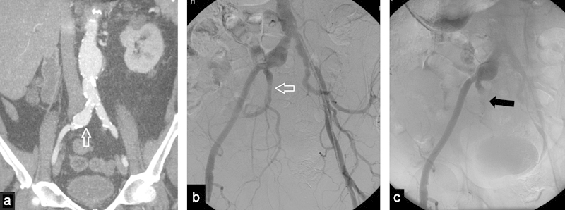
Fig. 5.

(a) Coronal image of a computed tomography with contrast, shows an abdominal aortic aneurysm with extension into the right common iliac artery (arrow). (b) Angiogram before embolization shows right common iliac artery aneurysm with patent right internal iliac artery (arrow). (c) Angiogram after internal iliac artery embolization with an AVP (arrow). Note very proximal embolization.
