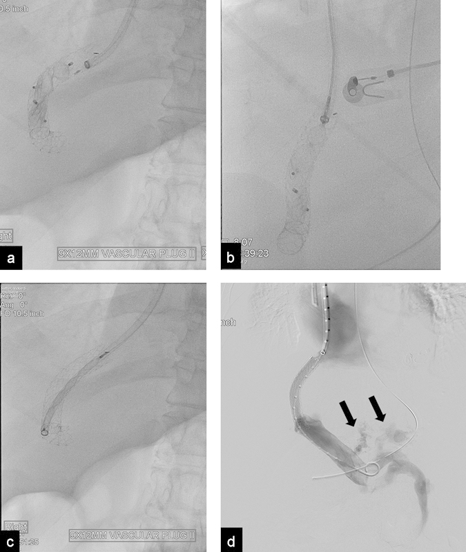
Fig. 8.

(a) Radiograph shows two AVPs used to close a TIPS in a patient with severe encephalopathy. Patient developed severe variceal bleeding after shunt closure. (b) Radiograph shows the proximal end of the plug being collapsed after it was grabbed with a snare. (c) Radiograph shows the plug completely collapsed inside a 10 Fr. sheath. (d) Portogram after plug retrieval and balloon dilatation of the shunt shows patent TIPS; the esophageal varices (arrows) were subsequently embolized.
