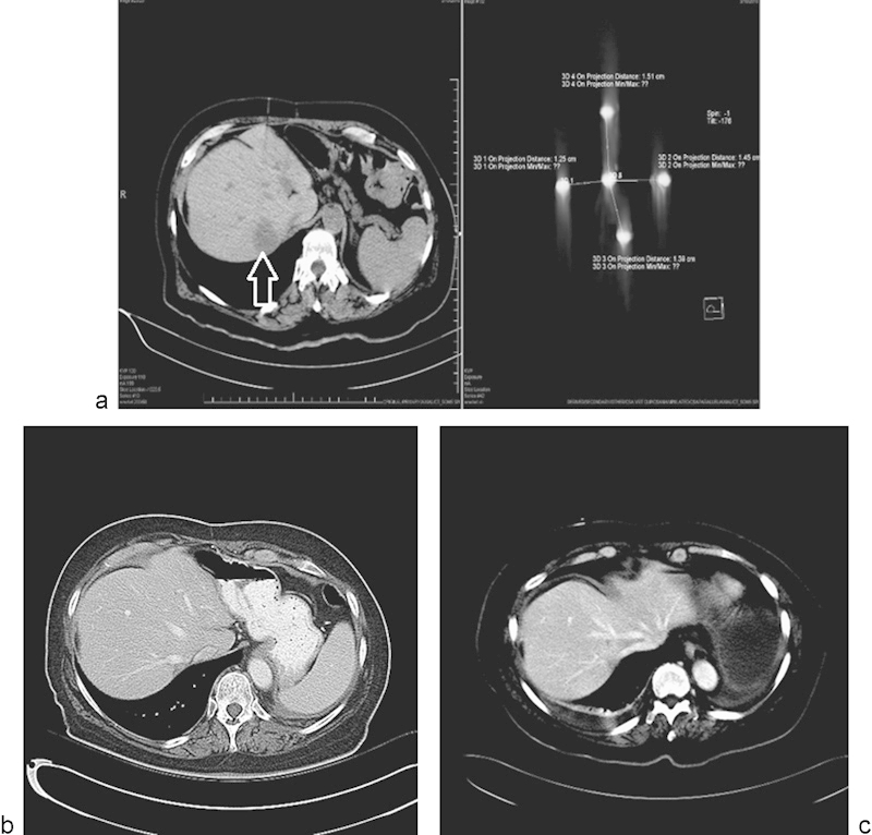
Fig. 4.

Single metastatic focus in a lung cancer patient treated with irreversible electroporation. (a) CT scan demonstrating the metastatic focus in the liver (arrow), and placement of the five probes used to treat the lesion. (b and c) Follow-up CT scan performed 8 and 12 months posttreatment, respectively, demonstrating no evidence for recurrent disease.
