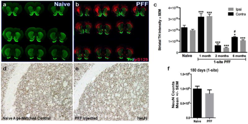
Figure 6. Bilateral α-syn pathology in the striatum ultimately results in reduced striatal innervation, but not neuronal cell death.

(a, b). Representative images of serial coronal sections proceeding from rostral to caudal, left to right, top to bottom, illustrating the pSyn immunoreactivity (red) and TH intensity (green) 6-months post injection (pi) in a naïve control rat (a) and a α-syn PFF 1-site rat. (c). Using near infrared integrated intensity measurements, we evaluated the extent of dopamine denervation in the striatum resulting from 1-site PFF injected animals at 30d, 60d and 180d and compared to naïve control. In PFF injected rats there was a significant bilateral increase in TH intensity at 30d pi followed by a significant bilateral reduction in the TH intensity at 60d and 180d compared to naïve animals. *** p < 0.0001 compared to naïve, * p<0.01 compared to naive, # p<0.03 compared to either ipsi or contra striatum at 2 months. (d, e). To determine whether pSYN accumulation in the striatum results in neuronal cell death, we utilized stereology to determine the total neuronal population (NeuN) throughout the rostro-caudal striatum. NeuN staining showed no significant differences between the naïve-age-matched striatum (d) and the PFF-injected striatum (e). (f). Stereological quantification of NeuN counts in the striatum (ipsilateral to injection site) 6 months post-PFF injection (1-site) compared to naïve control. Data represent mean +/−SEM. Abbreviations: PFF= pre-formed α-syn fibrils, TH=tyrosine hydroxylase, pS129= α-syn phosphorylation at serine 129, contra=contralateral hemisphere (relative to injection site), ipsi=ipsilateral hemisphere (relative to injection site, NeuN=neuronal nuclei.
