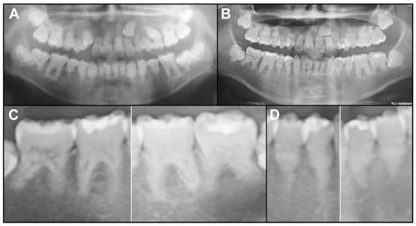
Fig. 2.
Panoramic radiographs. A, Panoramic radiograph at age 14 years. B, Panoramic radiograph at age 16 years. All 32 permanent teeth were visible, although the left maxillary cuspid (tooth #11) was impacted, with retention of tooth C. The tooth roots were shortened and misshaped. Pulp stones and obliterated pulp chambers are observed throughout. The enamel layer contrasted well with dentin in most teeth. Subtle radiolucencies were associated with some mandibular roots, although these teeth were vital and asymptomatic. C, Details of mandibular molars. Some roots (particularly #30 and 31) showed signs of external root resorption. D, Details of mandibular bicuspids. Single-rooted teeth exhibited midroot bulges with apical thinning.

