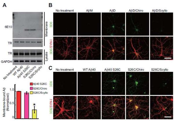
Figure 2. Prevention of Aβ oligomer binding to the surface of hippocampal neurons by scyllo-inositol.
(A) Representative anti-Aβ Western blot showing streptavidin-precipitated biotinylated Aβ on primary hippocampal neurons (DIV18) after various peptide treatments. Western blotting of transferrin receptor (TR), and TR and GAPDH in the cell lysate served as controls. Bars: mean levels of membrane-bound Aβ; cultures treated with (Aβ40 S26C)2 dimer only were normalized to 1 (*, p<0.01 by one-way ANOVA test). N = 4 independent experiments; error bars, s.e.m. (B , C) Confocal micrographs of hippocampal neurons (DIV21) show surface-bound Aβ detected by 3D6 (green) alone or co-labeled for synapsin I (SYN-I, red) after 3 hr treatment with Aβ monomers or Aβ dimers from AD-TBS (B) or pure, synthetic wt Aβ40 or (Aβ40 S26C)2 dimer (C) in the presence or absence of chiro- or scyllo-inositol (20 μM). Scale bar, 50 μm.

