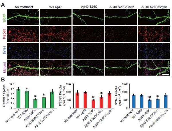
Figure 3. Scyllo-inositol inhibits.
A β oligomer-induced synaptic loss in primary hippocampal neurons. (A) Confocal images showing EGFP-labeled dendritic spines (green), endogenous PSD95 (red) and synapsin I (SYN-I, blue) of hippocampal neurons (DIV21) transfected with EGFP and exposed for 1 d to synthetic wt Aβ40 or (Aβ40 S26C)2 dimer with or without co-administration of chiro-inositol or scyllo-inositol (20 μM). Scale bar, 10 μm. (B) Bars: mean density of dendritic spines and PSD95/SYN-I puncta under different conditions of treatment. * significantly different from neurons without treatment (p < 0.05 by one-way ANOVA test). Fifteen cells per condition were analyzed. Error bars, s.e.m.

