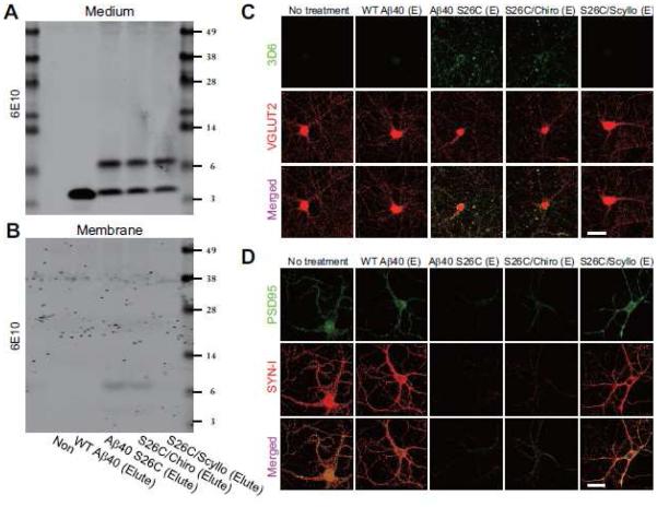
Figure 5. Pre-incubation of scyllo-inositol with soluble Aβ oligomers prevents their binding to neurons and synaptotoxic effects.
Representative blots showing eluted A β in culture medium (A) and bound on membrane (B) of hippocampal neurons (DIV18) under different treatments. (C) Confocal images show surface-labeled Aβ (detected by 3D6, green), and total VGLUT2 (red) of hippocampal neurons (DIV18) after 12 hr treatment with eluted wt Aβ40 or (Aβ40 S26C)2 dimer, with or without co-administration of chiro-or scyllo-inositol (20 μM). Scale bar, 20 μm. (D) Confocal images showing endogenous PSD95 (green) and synapsin I (SYN-I, red) in hippocampal neurons (DIV18) after 1 d treatment with eluted synthetic wt Aβ40 or (Aβ40 S26C)2 dimer, with or without co-administration of chiro-or scyllo-inositol (20 μM). Scale bar, 20 μm.

