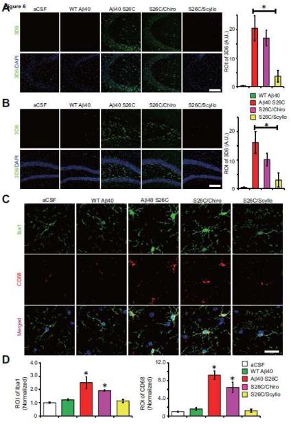
Figure 6. Prevention of Aβ oligomer binding to the neuronal surface and Aβ oligomer-induced activation of microglia in vivo by scyllo-inositol.
Confocal micrographs showing surface membrane-bound Aβ detected by 3D6 (green), and DAPI (blue) in CA3 (A) and DG (B) of hippocampus 1 day after ICV injection with synthetic wt Aβ40 or (Aβ40 S26C)2 dimer in the presence or absence of chiro- or scyllo-inositol (20 μM). Scale bar, 100 μm. Bars: mean intensity of 3D6 labeling under indicated conditions. *significantly different from mice injected with wt Aβ40 (p < 0.05, one-way ANOVA test). Eighteen slices per condition were analyzed. Error bars, s.e.m. (C) Confocal images showing the pattern of microglia labeled by Iba1 (green) and CD68 (red) in CA3 of hippocampus 4 day after ICV injection with synthetic wt Aβ40 or (Aβ40 S26C)2 dimer in the presence or absence of chiro- or Scyllo-inositol (20 μM). Scale bar, 20 μm. (D) Bars: mean intensity of Iba1 and CD68 under different conditions. * significantly different mice injected with aCSF (p < 0.05 by one-way ANOVA test). Fifteen slices per condition were analyzed. Error bars, s.e.m.

