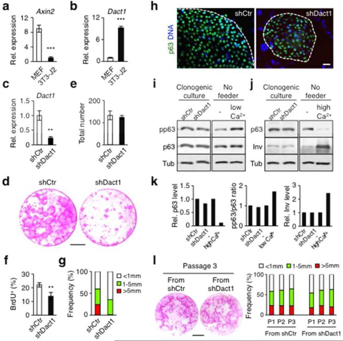
Figure 1. Suppression of Dact1 in 3T3-J2 cells reduces clonogenic expansion of HPEKs.
(a) Relative expression of Axin2. (b) Expression of Dact1 in 3T3-J2 cells. (c) Suppression of Dact1 by shRNA (shDact1) in 3T3-J2 cells. shCtr, control shRNA. (d) Rhodamine staining of epidermal clones grown for 14 days. Bar=10 mm. (e) Total number of epidermal clones at day 14. (f) Proliferation of keratinocytes in clonogenic culture at day 7. (g) Size distribution of epidermal clones at day 14. (h) Expression of p63 in representative epidermal clones. Bar=20 μm. (i and j) Western blot of keratinocytes from clonogenic cultures. HPEKs stimulated with low (i: 0.3 mM) or high (j: 1.3 mM) calcium, as positive controls. (k) Quantification of i and j. (l) Serial culture of equal plated numbers of keratinocytes, grown for 14 days with γ-irradiated fresh 3T3-J2 cells. Left: rhodamine staining at passage (P) 3. Right: size distribution of epidermal clones over multiple passages. Bar=10 mm. Data shown are mean±SEM (n=3). **P<0.01; ***P<0.001.

