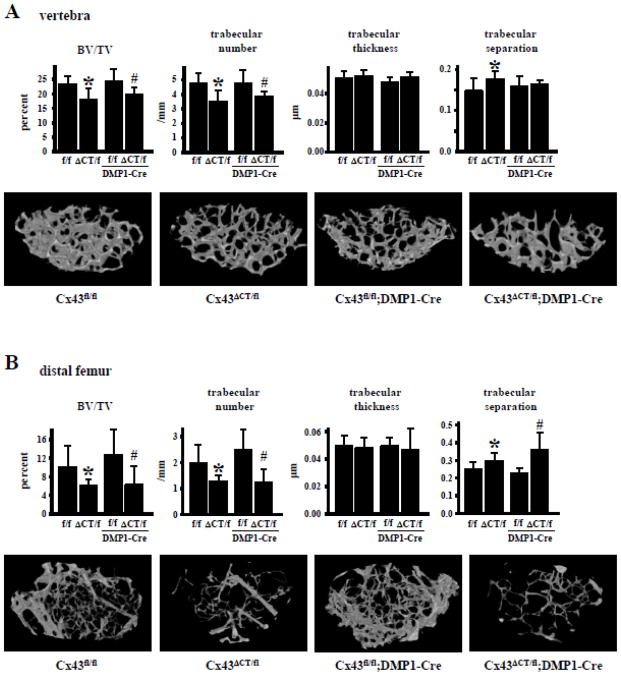
Fig. 2. Absence of CT domain of Cx43 results in low cancellous bone volume.
Cancellous bone microarchitecture was assessed in Cx43fl/fl, Cx43ΔCT/fl, Cx43fl/fl;DMP1-8kb-Cre, and Cx43ΔCT/fl;DMP1-8kb-Cre mice at 4.5 months of age by μCT in (A) L4 vertebrae and (B) distal femora. Representative reconstructed 3D μCT images are shown. Bars represent mean ± s.d., n=7–12. *p<0.05 versus Cx43fl/fl mice, and #p<0.05 versus Cx43fl/fl;DMP1-8kb-Cre mice by two-way ANOVA.

