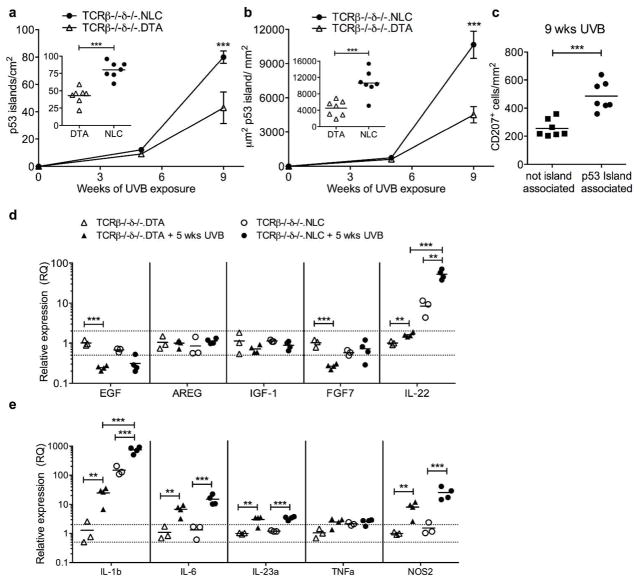
Figure 4. LC facilitation of p53 island growth is T cell independent.
Mutant KC p53 island density (a) and area (b) were quantified in epidermal sheets prepared from TCRβ−/−δ−/−.NLC and TCRβ−/−δ−/−.DTA mice following 5 or 9 wks UVB exposure (400 J/m2, 3x/wk). Inset graphs show distribution at 9 wks; each symbol represents one mouse. LC-intact TCRβ−/−δ−/−.NLC mice have increased p53 island density and area compared to LC-deficient TCRβ−/−δ−/−.DTA mice following 9wks UVB; ***P<0.001. (c) CD207+ LC density is increased in association with p53 islands. (d, e) Epidermal cells prepared from untreated vs 5 wk chronic UVB treated TCRβ−/−δ−/−.NLC and TCRβ−/−δ−/−.DTA mice were examined for changes in gene expression (relative to untreated TCRβ−/−δ−/−.DTA) by qRT-PCR; each symbol represents one mouse,**P<0.01, ***P<0.001, Holm-Sidak correction for multiple comparisons.

