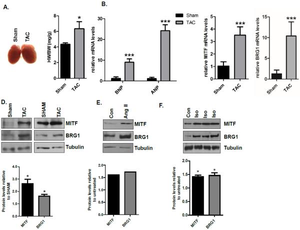
Fig.1. MITF and BRG1 mRNA and protein levels increase after transverse aortic constriction (TAC).
(A) Representative images of hearts and heart weight/body weight (HW/BW) in sham and TAC mice 2 weeks after surgery. (B) Relative BNP and ANP expression levels were measured by quantitative RT- PCR (qRT – PCR) from left ventricles of sham and TAC mice described in (A). ANP and BNP levels were normalized to 18s rRNA. (C) MITF and BRG1 expression levels were assessed by qRT-PCR as in B. For experiments shown in (A), (B), and (C), the sample size was N=3 for sham, N=4 for TAC. Standard error bars are shown (* p<0.05, **p<0.01, and *** p<0.005). (D) Whole cell lysates from sham and TAC mice described in A were run on an SDS-polyacrylamide gel and immunoblotted with MITF and BRG1 antibodies. Tubulin was used as a loading control. Each blot is from an independent experiment in which cardiomyocytes were obtained from three to five mice. (E) Whole cell lysates from control and angiotensin II (200nM) treated primary adult mouse cardiomyocytes were prepared six hours after treatment, run on an SDS-polyacrylamide gel and immunoblotted with MITF and BRG1 antibodies. Tubulin was used as a loading control. The blot is from an experiment in which cardiomyocytes were obtained from four mice. (F) Whole cell lysates from control and isoproterenol (10μM) treated primary adult mouse cardiomyocytes were prepared six hours after treatment, run on an SDS-polyacrylamide gel, and immunoblotted with MITF and BRG1 antibodies. Tubulin was used as a loading control. Each lane on the gel contains whole cell lysate isolated from cardiomyocytes that were isolated from one mouse. Band densities were quantified using Image J software and shown in the graphs below each blot.

