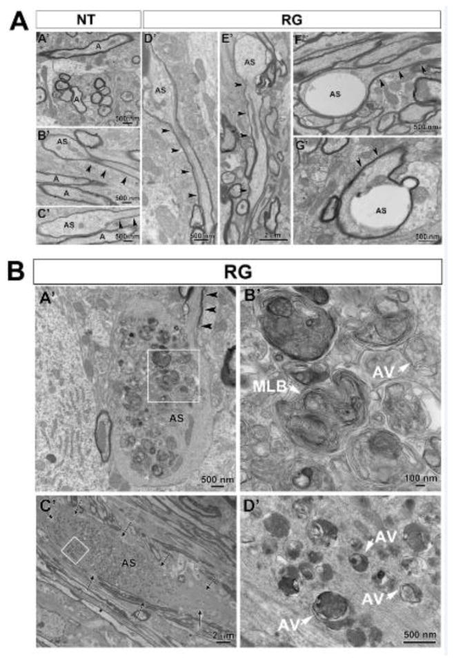
FIGURE 6.
Transmission electron microscopic analysis of axonal spheroids in the medial forebrain bundle (MFB) in the hLRRK2(R1441G) BAC transgenic mice and non-transgenic litter-mate controls. (A) A representative electron micrograph (A′) shows the normal ultrastructure of axons (A) in the MFB in non-transgenic mice (NT). In B′ and C′ examples are shown of small axonal spheroids (AS) such as were occasionally observed in thinly myelinated axons in the NT mice. Thus ultrastructural analysis confirms the simple, oval morphology observed by TH immunohistochemistry (Figure 2C) and by Tau-tdTomato anterograde labeling (Figure 3A). Note that the cytoskeleton of the normal portion of the axon (black arrowheads) becomes disrupted as it enters the spheroid. A similar type of simple spheroid morphology with disruption of cytoskeleton was observed in the hLRRK2(R1441G) BAC transgenic mice (RG) (D′ and E′). The RG mice also demonstrated another type of spheroid which was relatively devoid of cellular organelles and cytoskeletal elements, leaving large, clear electron-lucent swellings (F′ and G′). (B) Unique to the RG mice were giant spheroids that contained large numbers of autophagic vacuoles. In A′ a thinly myelinated axon (arrowheads) with well-organized cytoskeletal elements leads into a large spheroid that lacks myelin, contains disorganized cytoskeletal structures and is packed with autophagic vacuoles. The region in the white box is shown at higher magnification in B′. Numerous vacuoles with double membranes, characteristic of autophagic vacuoles (AV), are observed. Interspersed among them are numerous multilamellar bodies (MLB), frequently described in association with classic autophagic vacuoles (Hariri et al., 2000; Hornung et al., 1989; Nixon et al., 2005). In C′ an unmyelinated large spheroid (delineated by arrows) is also packed with numerous autophagic vacuoles. The region in the white box is shown at higher magnification in D′. A, axon; AS, axonal spheroid. Arrowheads always indicate the intact portion of the axon containing the axonal spheroids.

