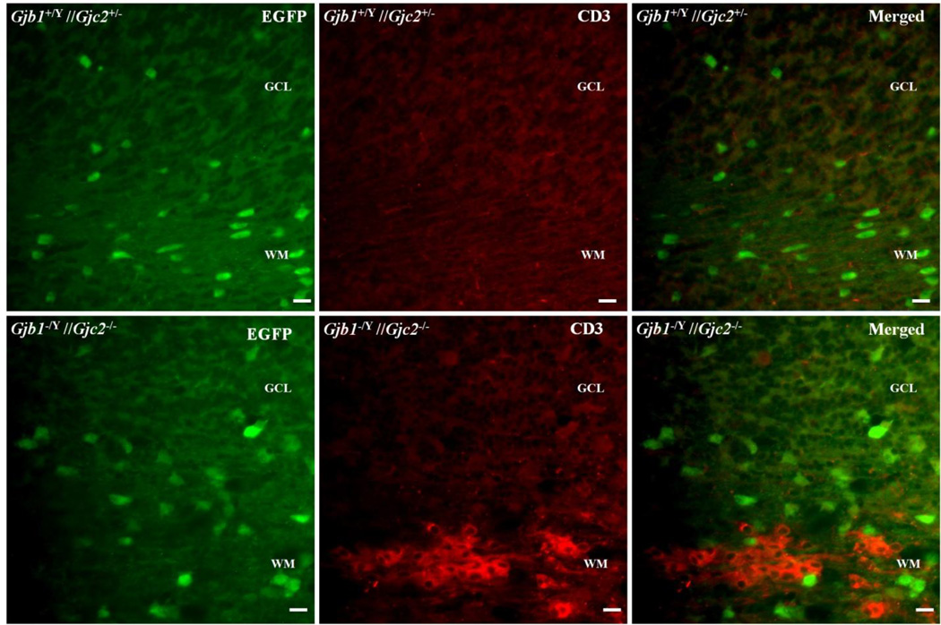
Figure 4. Microglial and astrocytic responses in Gjb1−/Y//Gjc2−/− cerebella.
These are digital images of sections from the cerebella from P29 Gjb1−/Y//Gjc2−/− mice and their littermate controls (Gjb1+/Y//Gjc2+/−), immunostained for Iba1 or GFAP. The molecular layer (ML), granular cell layer (GCL), and white matter (WM) are labeled. The upper panels show activated microglia in the WM of a Gjb1−/Y//Gjc2−/− cerebellum but not a littermate control. The lower panels show increased GFAP staining in the WM of a Gjb1−/Y//Gjc2−/− cerebellum compared to the control. Scale bars: 10 µm.

