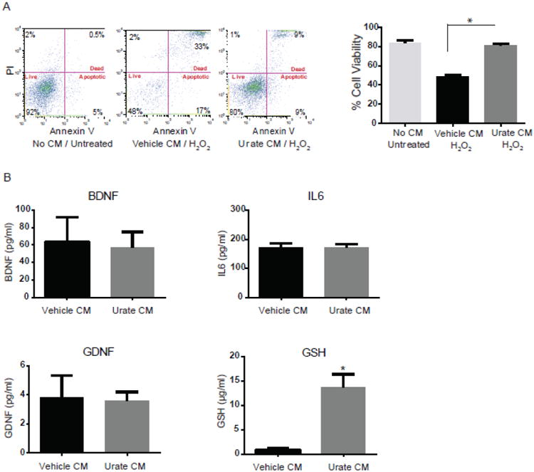
Figure 1. Marked elevation in GSH release from astrocytes treated with urate.

(A) Conditioned medium (CM) from urate-treated astrocytes protects dopaminergic cells from H2O2-induced cell death. Representative graphs of FACS analysis show cell viability using propidium iodide (PI)/Annexin V staining. Percentages of PI+/AnnexinV+ (dead), PI-/AnnexinV+ (apoptotic) and PI–/AnnexinV– (vital) staining are shown for untreated MES 23.5 cells, or those treated with CM from vehicle- or urate (100 μM)-treated astrocytes a day before and during 200μM H2O2 treatment for 24h. (B) Targeted screening of several neurotrophic factors in the CM. There was no change detected in levels of BDNF, GDNF and IL6 factors in the urate versus vehicle CM. GSH content was significantly increased in CM from urate- (versus vehicle-) treated astrocyte. *denotes p value < 0.001; (n=4 independent experiments).
