
Figure 3.
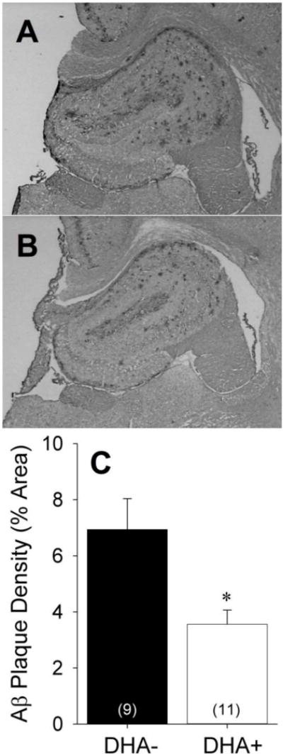
Representative hippocampal sections from APP/PS1 rats maintained on A) DHA-depleted (DHA−) or B) DHA-supplemented (DHA+) diets labeled with the anti-Aβ antibody DAE. C) Aβ plaque density in the DHA− and DHA+ groups expressed as percentage of hippocampal area. *p<0.05 vs. DHA− group.