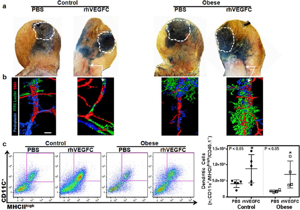
Figure 4. Subcutaneous injection of rhVEGF-C in ear skin improves lymphatic function and decreases contact hypersensitivity in both lean and obese mice.
a. Evans blue lymphangiography of control and obese mouse ears 3 days after DNFB challenge. Mice were injected in the base of the ear with either PBS or rhVEGF-C subcutaneously once daily for 7 days and then challenged with DNFB. Subsequently, PBS and rhVEGF-C injections were continued for an additional 3 days post-challenge after which lymphangiography was performed. White dashed line area represents Evans blue injection site. Arrows show dye in collecting lymphatic vessels at the base of the ear in control and obese mice injected with rhVEGF-C.
b. Representative ear whole mount images of FITC lectin lymphangiography of control and obese mouse ears 3 days following DNFB challenge in animals treated with daily PBS or rhVEGF-C injections for 10 days as outlined above. White arrows show functional lymphatic vessels containing intraluminal tomato lectin stain (green) in rhVEGF-C treated lean and obese mice groups. Scale bar=50µm.
c. Left panel: Representative flow cytometry plots of dendritic cells (CD11c+/MHCIIhigh/CD45.1+) harvested from cervical lymph nodes draining the ipsilateral ears of DNFB challenged control and obese mice treated with daily ear subcutaneous PBS or rhVEGF-C injections (7 days before and 3 days after DNFB challenge). Purple box represents positive cells. Right panel: Quantification of the migrated DCs from ear skin to draining lymph nodes in various groups (n=5 animals/group; *p<0.05). Note increased DC trafficking in lean and obese mice injected with rhVEGF-C.

