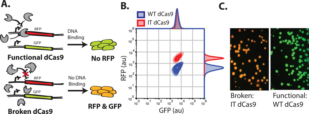
Figure 3.
Screen for functional Cas9s. (A) Schematic representation of the screen. (B) Flow cytometry data of the functional positive (WT dCas9) control in blue and negative 'Inactive Truncation' Cas9 (IT dCas9) control in Red, IT dCas9 contains only the C-terminal 250 amino acids. Both controls contain the sgRNA plasmid targeting RFP for repression. Samples were grown overnight in rich induction media. (C) Colony fluorescence of the functional (WT dCas9) and 'Broken' negative (IT dCas9) controls.

