Fig. 2.

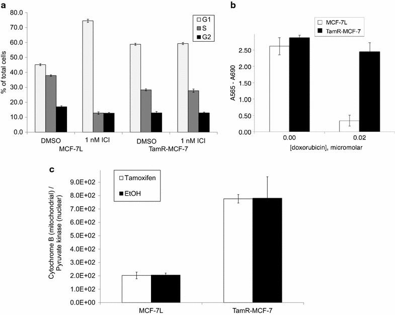
Tamoxifen resistant breast cancer cells have elevated mitochondrial DNA and cross resistance to ICI and doxorubicin. a MCF-7L and TamR-MCF-7 cells were treated for 2 days in 1 nM ICI or vehicle control (DMSO), then harvested and fixed in ethanol. Cell cycle phase distribution was determined by flow cytometric measurement of propidium iodide staining per cell. The average percentage of G1, S and G2/M cells are shown; error bars indicate one standard deviation (n = 3). b MCF-7L and TamR-MCF-7 cells were treated ±0.02 mm doxorubicin. After 4 days, total cellular mass was measured by sulforhodamine B staining. The average SRB staining is shown; error bars indicate one standard deviation (n = 6). c MCF-7L and TamR-MCF-7 cells were treated for 2 days with 10 nm 4-hydroxytamoxifen or control (EtOH). Genomic DNA was extracted and used as template for detection of cytochrome B DNA on the mitochondrial genome and pyruvate kinase DNA on the nuclear genome. The average ratio of cytochrome B to pyruvate kinase is shown; error bars indicate one standard deviation (n = 3)
