Fig. 3.

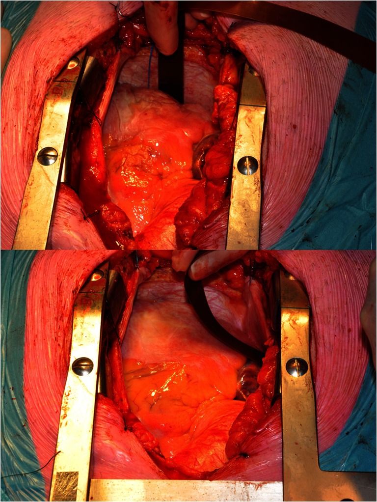
The electrode array placed between the pulmonary veins in the oblique sinus (top) and on the right atrial wall (bottom) during recording of epicardial electrograms

The electrode array placed between the pulmonary veins in the oblique sinus (top) and on the right atrial wall (bottom) during recording of epicardial electrograms