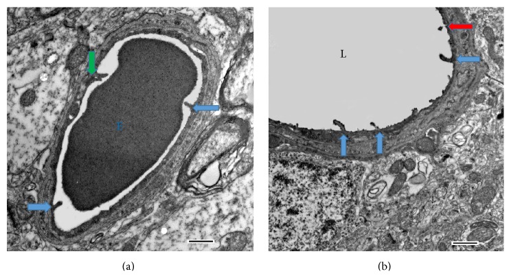
Figure 1.
The microvilli in the rat capillary: electron microphotography. (a) An erythrocyte (E) inside the capillary; the microvilli (arrows) produce visible invaginations on the erythrocyte wall. (b) A larger blood vessel. L: the lumen of the vessel. The microvilli are marked with arrows; green arrow: double microvilli (see the text); red arrow: a radially cut microvillus, with axial symmetry. Scale bar: 500 nm.

