. 2015 Sep;48(9):495–500. doi: 10.5483/BMBRep.2015.48.9.259

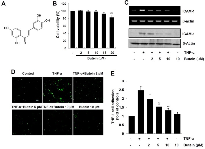
Fig. 1. Effect of butein on TNF-α-induced expression of ICAM-1 and monocyte adhesion in HaCaT cells. (A) Chemical structure of butein. (B) HaCaT cells were incubated with various concentrations of butein for 24, and then cell viability was evaluated by MTT assay. The results are expressed as mean ±SD of three independent experiments. Statistical significance: ***P < 0.001 compared to control group. (C) Cells pretreated with 2, 5 and 10 μM butein for 4 h were exposed to 10 ng/ml TNF-α for 1 h (for RNA), or 12 h (for protein). Total RNA and protein were analyzed by RTPCR (upper panel), and Western blotting (bottom panel), respectively. (D) HaCaT cells were incubated with 2, 5 and 10 μM butein for 4 h, and then exposed to 10 ng/ml TNF-α for 12 h. Calcein-AM-labeled THP-1 monocytes were added, and incubated with HaCaT cells for 1 h. Microscopic images were obtained using a fluorescence microscopy (scale bar=50 μm). (E) Calcein-AM fluorescent intensity was quantified using a fluorescence plate reader. The results are expressed as mean ± SD of three independent experiments. Statistical significance: **P < 0.01 compared to TNF-α alone.

Fig. 1.