. 2015 Sep;48(9):531–536. doi: 10.5483/BMBRep.2015.48.9.117

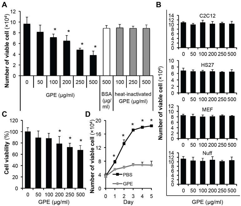
Fig. 1. GPE induces the cell death of 5637 cells. (A) 5637 cells were seeded in 24-well cell culture plates, at a density of 2 × 10⁴ cells/well. Next day, cells were treated with indicated concentrations of GPE, BSA or heat-inactivated GPE, and further incubated for 48 h. Cells were then trypsinized, and the number of viable cells was counted. *P < 0.05, compared with no GPE treatment. (B) Cells were seeded in 24-well cell culture plates at a density of 2 × 10⁴ cells/well. Next day, cells were treated with indicated concentrations of GPE and further incubated for 48 h. Cells were trypsinized and the number of viable cells counted. (C) 5637 cells were treated with indicated concentrations of GPE for 48 h and the MTT assay was performed, as described in Materials and Methods. *P < 0.05, compared with no GPE treatment. (D) The number of viable 5637 cells treated with control buffer (PBS) or GPE (500 μg/ml) were counted daily. *P < 0.05, compared with respective day control.

Fig. 1.