Figure 3.

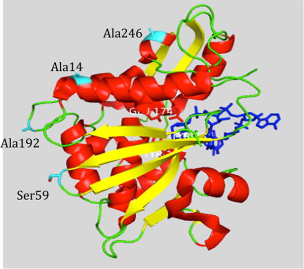
Crystal structure of saporin L3. The structure is shown in cartoon representation. The RNA ligand is colored blue. The residues colored cyan are targets for mutation to cysteine. The structure is from Protein Data Bank entry 3HIW.

Crystal structure of saporin L3. The structure is shown in cartoon representation. The RNA ligand is colored blue. The residues colored cyan are targets for mutation to cysteine. The structure is from Protein Data Bank entry 3HIW.