
Figure 6. BJAB K20 cells poorly metabolize protected sialic acids.
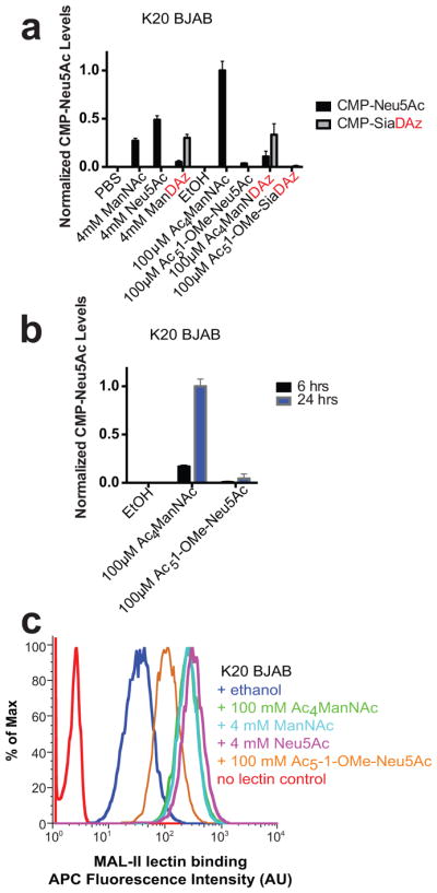
(a) BJAB K20 cells were cultured with indicated molecules for 24 h and intracellular levels of CMP-sialic acids were quantified by HPAEC-UV. Analysis was performed in biological duplicate with error bars showing standard deviation of the mean. (b) BJAB K20 cells were cultured with 100 μM Ac4ManNAc or 100 μM Ac5-1-OMe-Neu5Ac for 6 or 24 h. Intracellular CMP-Neu5Ac was quantified by HPAEC-UV. Analysis was performed in biological duplicate with error bars showing standard deviation of the mean. (c) BJAB K20 cells were cultured with indicated molecules for 48 h. Cell surface α2–3-linked sialic acid was measured by flow cytometry using biotinylated MAL II lectin and APC-streptavidin. Data presented are a single trial representative of replicate experiments.