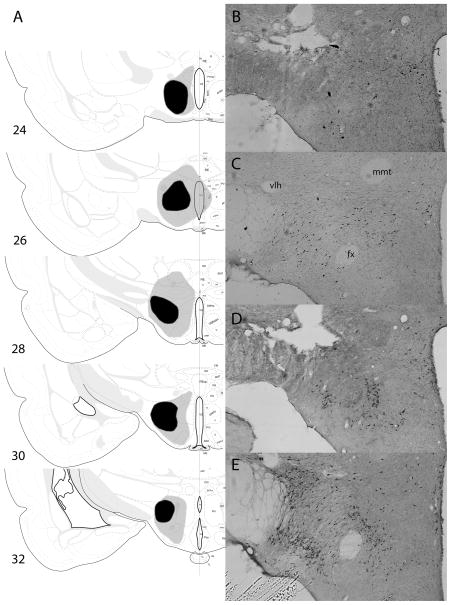
Fig. 3.
(A) Drawings of the largest (gray) and smallest (black) accepted lesions of lateral hypothalamus (LH), based on Nissl-stained sections, in experiment 2. All lesions are shown as if they were made in the same hemisphere. (B–E) Sample lesioned (B and D) and unlesioned (C and E) sections, stained for orexin (B and C) or melanin-concentrating hormone (MCH; D and E). The images all came from the same rat, with those shown in (C and E) reversed left-to-right to portray all sections in the same orientation, matching (A). The images in (B and C) are from the same section, and the images in (D and E) are from the same section, adjacent to the section displayed in (B and D). fx, fornix; mmt, mammillothalamic tract; vlt, ventrolateral hypothalamic tract. Drawings of brain sections in (A) are from Swanson (1998); used by permission of Elsevier.

