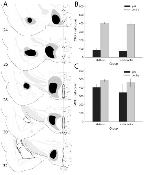
Fig. 9.
(A) Drawings of the largest (gray) and smallest (black) accepted lesions of amygdala central nucleus (CeA) and lateral hypothalamus (LH), based on Nissl-stained sections, in experiment 3. All lesions are shown in the same hemisphere, regardless of whether they were made in the right or left hemisphere. (B and C) Mean ± SEM counts of ORX+ and MCH+, respectively, cells in the hypothalamus of rats that were trained in the shift condition (Table 1). The rats in the shift-Contra group received an orexin-saporin lesion of the LH in one hemisphere and an ibotenic acid lesion of the CeA in the other, whereas rats in the shift-Uni group received only a unilateral LH lesion. The black bars (Ipsi) refer to counts in the lesioned hemisphere and the gray bars (Contra) refer to counts in the undisturbed hemisphere. Drawings of brain sections in (A) are from Swanson (1998); used by permission of Elsevier.

