
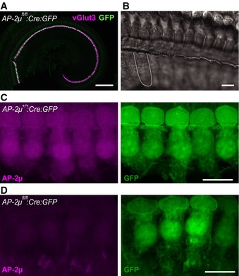
Figure 1. Hair cell‐specific disruption of AP‐2μ.
-
ACre recombination in IHCs and outer hair cells (OHCs) by transgenic Cre expression was demonstrated by a GFP reporter and used for inactivation of the AP‐2μ gene. Most if not all IHCs (identified by vGlut3 immunofluorescence, magenta) in an AP‐2μ fl/fl :Cre:GFP p14 organ of Corti are GFP positive (green). GFP fluorescence is weak in OHCs peripheral to IHCs and absent from SGNs (see also Fig EV1). Scale bar, 200 μm.
-
BDifferential interference contrast images indicate normal gross morphology of the p14 IHC hair bundle and basolateral pole. The dashed line indicates the approximate outline of an IHC. Scale bar, 20 μm.
-
C, DMaximum intensity projections of representative confocal sections of AP‐2μ +/+ :Cre:GFP (C) and AP‐2μ fl/fl :Cre:GFP (D) IHCs immunolabeled for AP‐2μ (magenta, left) and GFP (green, right). Scale bars, 10 μm.