
Figure EV1. Generation of hair cell‐specific AP‐2μ knockout mice (AP‐2μ fl/fl :Cre).
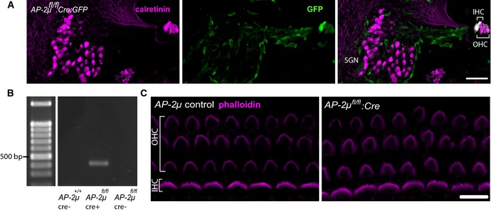
- Cryosectioned cochlea of a p7 AP‐2μ fl/fl :Cre mouse demonstrates VGLUT3‐driven Cre recombination in IHCs and OHCs (Jung et al, 2015) but not in SGNs, as reported by a floxed EGFP reporter (Nakamura et al, 2006). Immunohistochemical staining for calretinin (magenta), marking IHCs, OHCs, and SGNs and native EGFP fluorescence (green), indicating Cre recombination. In addition to EGFP signal in hair cells, some EGFP expression was found in capillaries and supporting cells. Scale bar, 50 μm.
- PCR of genomic DNA extracted from microdissected cochlea of p21 mice shows amplification of the Cre‐recombined region in Cre‐expressing AP‐2μfl/fl mice ((Kononenko et al, 2014), middle column), but not in AP‐2μ control: C57Bl/6 expressing Cre (left column) or AP‐2μ fl/fl mice lacking Cre (right column). Genomic DNA was isolated from whole cochleae of AP‐2μ fl/fl :Cre:GFP,AP‐2μ fl/fl, and AP‐2μ +/+:Cre mice with the nexttec™ kit (nexttec Biotechnologie GmbH, Germany). PCR was performed using DreamTaq DNA polymerase (Fermentas). The Cre‐recombinated excision of exon 2–3 in the Ap‐2μ gene was confirmed by using the forward primer TM330 GCTCTAAAGGTTATGCCTGGTGG and the reverse primer TM191 CCAAGGGACCTACAGGACTTC that detected a fragment of 404 bp (PCR at 58°C, 25 cycles) in cochlear DNA.
- Phalloidin immunofluorescence shows apparently intact stereociliar bundles (magenta) of IHCs (1 row) and OHCs (three rows) in acutely dissected apical coils of the cochlea from AP‐2μ fl/fl control and AP‐2μ fl/fl :Cre mice at p7. Scale bar, 10 μm.