
Figure EV5. AAV2/1 gene replacement of AP‐2μ restores near‐normal otoferlin protein levels in IHCs of AP‐2μ fl/fl :Cre mouse.
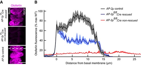
- Maximum projections of confocal sections of otoferlin‐immunolabeled organs of Corti of p14–17 AP‐2μ fl/fl :Cre rescued (AAV‐AP‐2μ‐injected; upper panel), AP‐2μ fl/fl :Cre non‐rescued (middle panel), and AP‐2μ control (bottom panel) ears that were stained and imaged in parallel under identical conditions. Scale bar, 10 μm.
- Average line profiles along the long axis of the IHCs (white dotted line in the bottom panel of A) reveal an increased otoferlin immunofluorescence in rescued IHCs, in particular at the plasma membrane in the basal pole where normal protein levels are found. Data were normalized to maximum fluorescence of IHCs from AP‐2μ control ears, originate from n = 19 IHCs for AP‐2μ control, n = 12 IHCs for AP‐2μ fl/fl :Cre rescued, and n = 7 IHCs for AP‐2μ fl/fl :Cre non‐rescued and are presented as mean ± SEM. The basal pole of the IHCs points to the left in the images. IHCs from AP‐2μ fl/fl :Cre rescued and AP‐2μ control ears happened to extend at a steeper angle within the organ of Corti in this sample than usual, giving rise to the shorter profiles in the maximum projection.