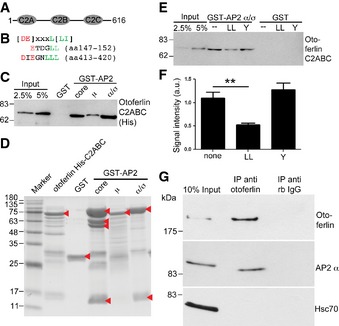
Otoferlin fragment used for binding assays.
Alignment of otoferlin dileucine‐containing motifs with consensus sequence.
Otoferlin directly associates with AP‐2. Bead‐bound GST‐AP‐2 core complex, GST‐AP‐2 α/σ hemicomplex, GST‐AP‐2μ, and GST were incubated with 225 nM of the His‐tagged otoferlin C2
ABC‐fragment. Samples were blotted and probed with His‐tag‐specific antibodies.
Coomassie gel of proteins used in (E, F). Arrow heads indicate purified proteins.
Dileucine motifs contribute to the binding between otoferlin and AP‐2. Bead‐bound GST‐AP‐2 α/σ hemicomplex and GST were incubated with 225 nM His‐tagged otoferlin C2
ABC after pre‐incubation with a dileucine‐containing peptide (LL), a tyrosine‐based peptide (Y), or no peptide (none). Samples were blotted and probed for otoferlin.
Quantification of data shown in (E). Data are given as mean ± SEM. Two‐tailed unpaired Student's t‐test to compare control (no peptide) with LL‐peptide treated condition, **P < 0.01. (n = 4).
Otoferlin and AP‐2 form a complex in living cells. Lysates from HEK cells overexpressing full‐length otoferlin and AP‐2μ were immunoprecipitated using anti‐otoferlin antibodies or control immunoglobulins. Samples were blotted and probed for otoferlin, and AP‐2α and Hsc70.